BMS2625 Biometrics SX230 EMG Pre-Amplifier: Ultrasound Imaging Compatibility
- Subject Code :
BMS2625
Question 1
The SX230 is an electromyography (EMG) pre-amplifier from Biometrics that has been used in several
clinical and research studies. It has two flying wire leads for use with reusable or disposable surface
EMG electrodes.
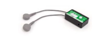
The sensor has the following specifications:
Two 4mm snap connectors on 100mm wires: use with reusable or disposable surface EMG
electrodes with 4mm studs
Gain: standard unit 1000
Bandwidth: 20 Hz - 460 Hz
Noise: < 5> Input Impedance: > 10,000,000 MOhms
Supply Voltage: +4.0 to +5.0 Vdc
Common Mode Rejection Rate (CMRR) @ 60 Hz (dB): > 96 dB (typically 110dB)
The block diagram in Figure 2 illustrates the main components of the Biometrics active EMG sensor.
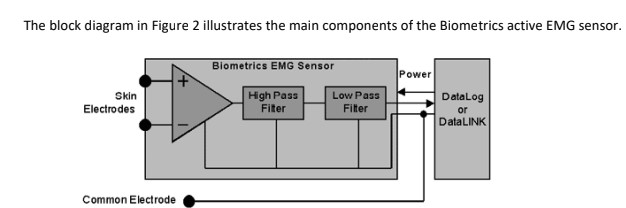
Main components are:
Differential electrodes to detect low level signals in a noisy environment.
A high-pass filter to remove DC offsets due to membrane potentials.
A low-pass filter to remove the unwanted frequencies above about 450Hz. It is very important
to remove these high frequencies when interfacing to a computer that is sampling the signal
since they would be converted to a lower frequency and mixed with the original signal.
A low noise instrumentation-amplifier front-end with a common-mode rejection ratio of
typically 110 dB.
Question 2
Ultrasound-based diagnostic imaging techniques are used to visualise subcutaneous body structures
including tendons, muscles, vessels and internal veins. For this question, you will have to:
a) Explain what are the differences among ultrasound A-mode, B-mode and M-mode. You should
discuss 2 different clinical applications for each mode and describe how the scan/image is
formed citing relevant literature sources. (weight 20%)
b) Identify relevant quality assurance protocols for testing of ultrasound scanners. (weight 10%)
WARNING
The work should be submitted via Turnitin a specific drop-box will be dedicated for this.
You are warned not to present somebody elses work as though it was your own, including another
students work. If you do this your work will be U graded which means it will be put under investigation
for academic dishonesty/plagiarism and, if confirmed given a P grade which will mean, at the very
least, you fail the module. However, you can easily avoid this action if you write up your work in your
own words and do your own analysis, graphs etc. Also, make sure that you properly cite any references
used.
Learning Outcomes
The following highlighted learning outcomes will be assessed:
LO1. Operate safely diagnostic equipment and troubleshoot a piece of equipment by using logical
deduction
LO2. Justify the choice of equipment with reference to its legal use in the clinical setting and explain in
detail how it works.
LO3. Evaluate the risks and benefits of using the equipment or technique.
LO4. Compare the quality assurance processes related to medical imaging.
Are you struggling to keep up with the demands of your academic journey? Don't worry, we've got your back! Exam Question Bank is your trusted partner in achieving academic excellence for all kind of technical and non-technical subjects. Our comprehensive range of academic services is designed to cater to students at every level. Whether you're a high school student, a college undergraduate, or pursuing advanced studies, we have the expertise and resources to support you.
To connect with expert and ask your query click here Exam Question Bank
